In a groundbreaking study, NIH-funded researchers have utilized advanced imaging techniques to uncover the structural foundations of memory formation in the mouse brain, offering new insights into how memories are formed at cellular and subcellular levels.
Introduction
Researchers at Scripps Research, supported by the National Institutes of Health (NIH), have employed cutting-edge imaging technologies to reconstruct the neural architecture underlying learning and memory in mice.
This study provides unprecedented insights into the flexibility of memory formation at both cellular and subcellular scales.
Methodology
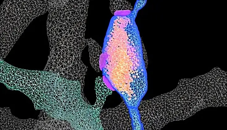
The research team combined advanced genetic tools, three-dimensional electron microscopy, and artificial intelligence algorithms to create detailed reconstructions of neural networks involved in learning.
Mice were subjected to a conditioning task, and their hippocampal regions were examined approximately one week later—a period post-initial memory encoding but prior to long-term storage.
Genetic labeling techniques enabled the identification of specific neurons activated during learning, facilitating precise analysis.
The Main Findings
Multi-Synaptic Boutons and Neural Flexibility
The study revealed that neurons associated with memory traces reorganized their connections through atypical structures known as multi-synaptic boutons.
In these configurations, a single neuron’s axon connects to multiple receiving neurons, potentially enhancing the flexibility of information coding within neural circuits.
Challenging Traditional Theories
Contrary to the longstanding theory that “neurons that fire together wire together,” the researchers found that neurons involved in the same memory trace did not preferentially connect with each other.
This finding suggests that memory formation may involve more complex and flexible neural interactions than previously thought.
Intracellular Reorganizations and Astrocyte Interactions
Neurons allocated to memory traces exhibited reorganization of intracellular structures responsible for energy production and communication support.
Additionally, these neurons showed enhanced interactions with astrocytes, a type of support cell, indicating a broader involvement of various cell types in memory formation.
Implications of the Study
These findings offer a comprehensive view of the structural hallmarks of memory formation in the hippocampus, a critical brain region for learning and memory.
Understanding the flexibility and complexity of these neural connections may provide insights into why memory and learning processes can malfunction, potentially informing strategies to address cognitive impairments.
Future Research Directions
The study opens avenues for further exploration, including:
-
Investigating whether similar mechanisms operate across different time points and neural circuits.
-
Examining the molecular composition of multi-synaptic boutons to determine their precise role in memory and other cognitive processes.
Continued research in these areas will be crucial to fully elucidate the mechanisms underlying memory formation and storage.
To Sum Up
This NIH-funded study provides significant insights into the structural features of memory formation, challenging traditional theories and highlighting the complexity of neural interactions.
The utilization of advanced imaging techniques has allowed for a more detailed understanding of how memories are formed and stored at the cellular level, paving the way for future research into cognitive processes and potential therapeutic interventions.
For a more in-depth exploration of this study and its implications, readers are encouraged to consult the original publication and related resources.
Sources: National Institutes of Health.